





Automatically translated by Google

Reviews are sourced from Google. Some reviews may be automatically translated and are displayed without editorial changes.
Automatically translated by Google

Automatically translated by Google

Automatically translated by Google

Automatically translated by Google

Automatically translated by Google

Automatically translated by Google

Automatically translated by Google

Automatically translated by Google

Automatically translated by Google
A prerequisite for implantation is a sufficient volume of bone in the jaw, making it possible to securely and stably anchor dental implants. However, if the residual bone volume is insufficient, bone transplantation (grafting) is necessary.
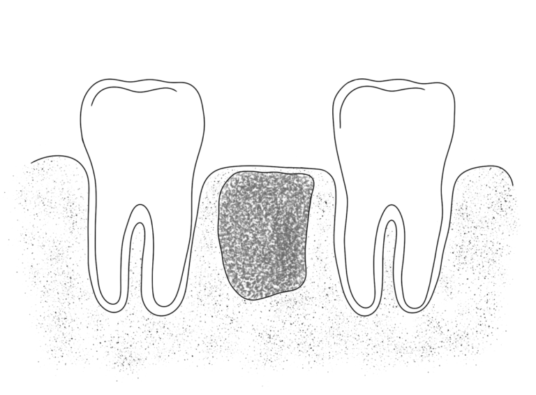
Materials for bone grafting or bone augmentation can be:
An autogenous, that is, one’s bone graft, is considered particularly suitable because it is well tolerated and is directly involved in the formation and development of new bone. In this method, the bone is taken from the mandible angle area. The disadvantage is that we can get a minimal amount of material, and this method requires additional surgical intervention.
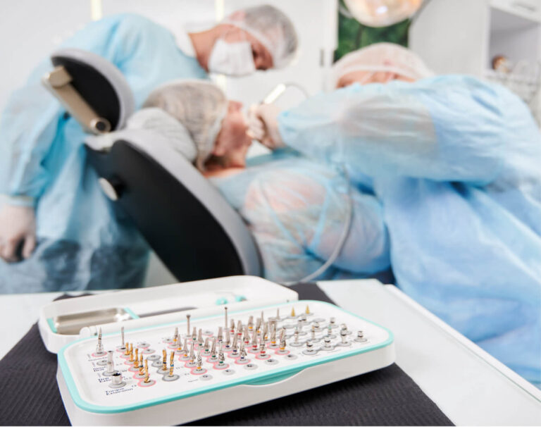
Allogeneic bone material. The advantages of allogeneic bone material are that it is available in any quantity and does not require additional surgery to harvest bone tissue. However, like any organ transplant, it can trigger an immune response in the recipient. Longer bone formation times should also be expected since allogeneic bone does not have the same favorable formation properties as autogenous material.
In recent years, good results have been achieved using xeno- and synthetic materials for human bone substitutes. Therefore, they are an excellent alternative to human bone.
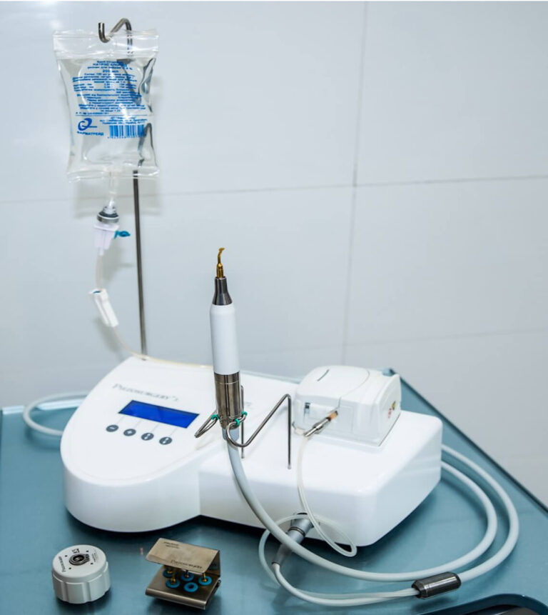
Piezoelectric bone surgery using a piezoelectric scaler is a method of performing surgical procedures in dentistry, such as tooth extraction, bone augmentation, and dental implantation. Under the influence of ultrasonic frequencies, even the hardest tissues are dissected, while soft tissues are not injured at all. The method allows surgical operations to be performed safely and minimally traumatic.
A centrifugal machine allows obtaining Platelet-Rich Fibrin (PRF), an autologous fibrin matrix produced from a patient’s platelet concentrate. The simplified manufacturing process conducted in the doctor’s office allows immediate generation of platelet-rich fibrin membrane that does not lose the potential to stimulate bone and soft tissue growth. The auto-biomaterial (PRF) main property is the slow release of growth factors over 7 days, speeding the wound healing process.
For example, if there is a residual ridge resorption, we use the osseodensification method applying Densah Bursversah instruments to perform sinus lifting or increase the alveolar bone volume. These instruments allow thickening of bone tissue, expand the alveolar ridge without bone grafting, or avoid sinus lift surgery.
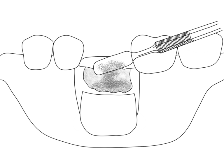
INLAY GRAFTING is used during tooth extraction. Bone material is placed in the extracted tooth’s socket to preserve the jaw bone volume and avoid bone resorption. This is a standard procedure for tooth extraction.
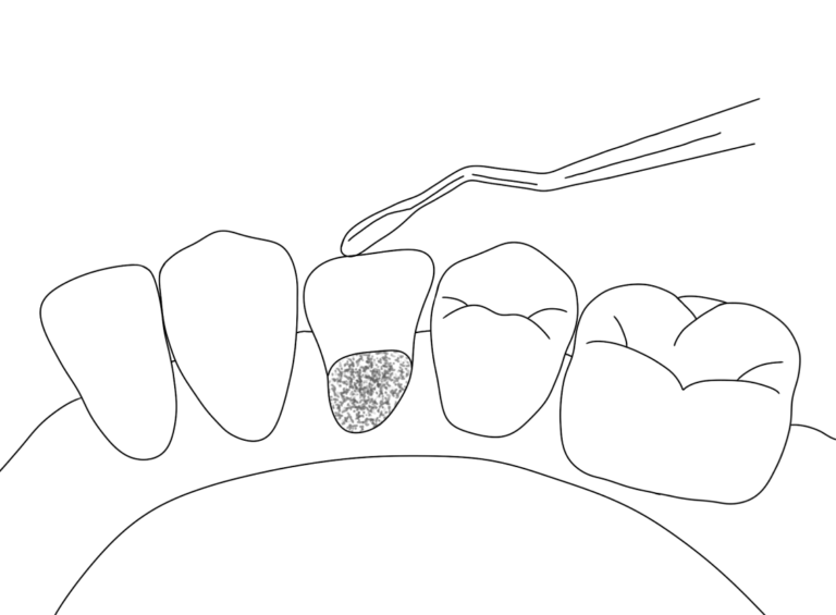
ONLAY GRAFTING. This procedure is used to restore the width and height of the residual bone. Bone material is placed on the jaw’s surface, or the jaw itself is split, and the gap between the bone fragments is filled with it.
SINUS GRAFTING. The sinus is the air space in the upper jaw (maxillary sinus) that can be used for bone grafting.
Today, bone grafting or bone augmentation has become a widespread procedure in dental surgery, which gives good results with minimum complications.
The reasons may be the following:
After tooth loss, an alveolar bone width decrease (sagittal type) tends to occur in the upper jaw, and an alveolar bone height decrease (vertical type) – in the lower jaw. The alveolar bone can shrink to a few millimeters. Prolonged use of removable dentures significantly increases alveolar bone atrophy.



 (580)
(580)
 (15)
(15)

There are several ways to rebuild bone loss.
The most reliable method is considered an increase with the help of the patient’s bone (from the donor site, for example, from the corner of the lower jaw, the bone tissue is carefully taken, placed in the future implantation site). This technique gives a positive result. You can also use bovine-derived xenogeneic materials. Such materials also give a positive result.
Sometimes we use synthetic materials to repair minor bone defects.
Depending on the size of the bone defect, the amount and type of bone graft material used, implantation can be performed 4-9 months after bone graft. For example, after open sinus lifting, we place implants 9 months later and 4 months later after alveolar ridge restoration using our autologous bone.
Xenomaterial and allografts are most often used to fill in the missing bone volume. These bone materials look like white crystals of different sizes and are stored in sterile packages.
The gold standard is autologous bone grafting – the patient’s bone. It is taken with a particular instrument from the corner of the lower jaw, chin, iliac or tibia. This type of bone gives the best result but is traumatic and more painful.
Before implantation, the dentist will always refer you for a CT scan.
When analysing the CT scan, the dentist will take measurements of the remaining bone and determine whether implantation is possible and whether bone grafting is necessary.





 Our team
 Reviews
 Blog
Blog News
News Partners
 FAQ
FAQ Vacancies
 Dental Consultation
 Tooth Decay Treatment
 Root canal treatment under a microscope
 Orthodontics
 Dental Implants
 Dental Prosthetics
 Professional Oral Hygiene
 Tooth Filling
 Periodontal disease treatment
 Aesthetic Dental Restoration
 Teeth extraction
 Surgical dentistry
 Sedation dentistry (sleep dentistry)