


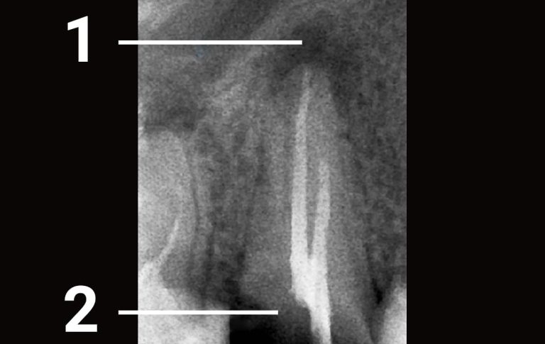
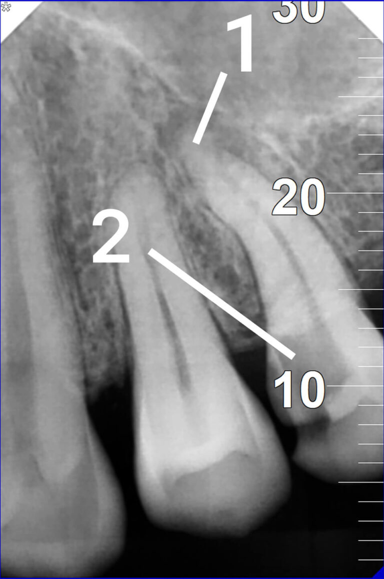
Before
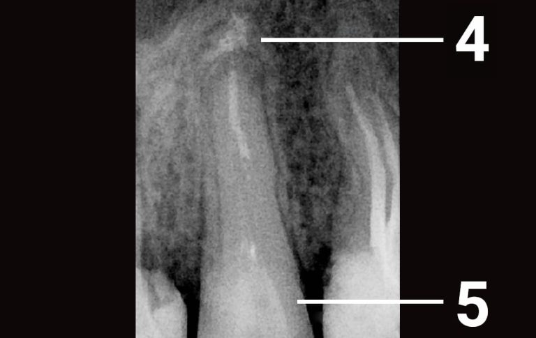
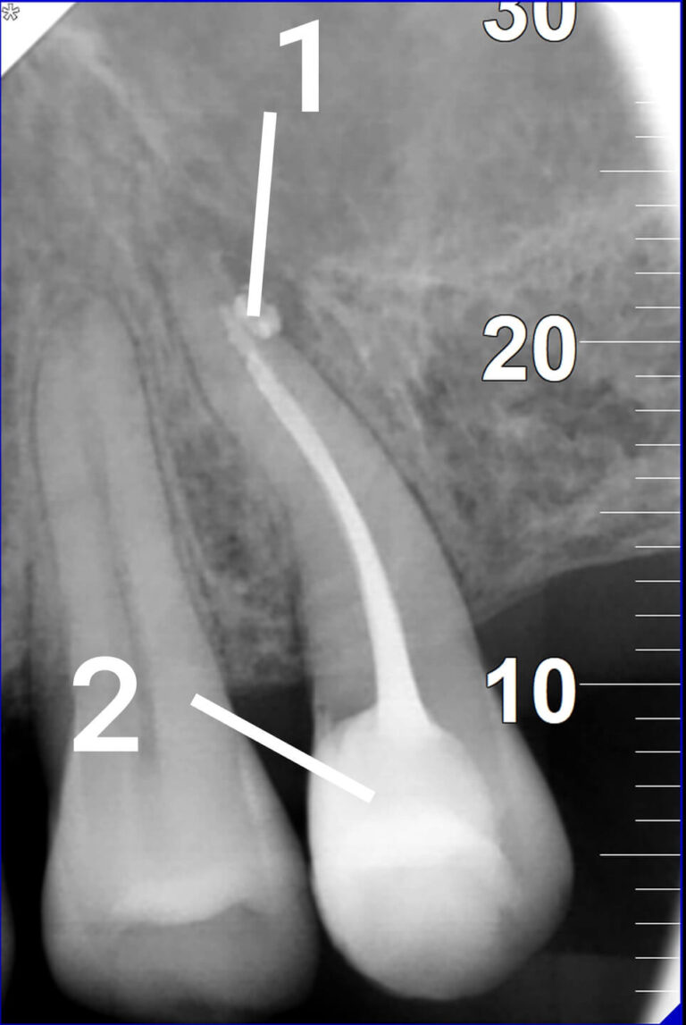
After

In brief: an X-ray revealed internal root resorption combined with asymptomatic apical periodontitis. The tooth was saved endodontically: the canal and the irregular resorption cavity were thoroughly cleaned and disinfected, then the defect was sealed (gutta-percha / bioceramic-MTA). At 8 months the follow-up X-ray showed bone recovery. Internal resorption is treated predictably when there is no root perforation.
Key facts
| Tooth | Lower jaw |
| Complaint | Intermittent sensitivity when chewing |
| Diagnosis | Asymptomatic apical periodontitis + internal root resorption |
| Method | Endodontics: cleaning and obturation of the canal and resorption area |
| Defect material | Bioceramic / MTA (for the irregular cavity) |
| Follow-up | At 8 months — bone recovery |
| Prognosis | Favourable when there is no perforation |
What is internal root resorption
Internal resorption is destruction of the hard tooth tissues from inside the root canal by specialised cells (odontoclasts) against a background of chronic pulp inflammation. The root walls thin out and an irregular, widened cavity forms within the canal. The condition is symptom-free for a long time; sometimes the only signs are mild sensitivity or a pinkish tint of the crown (the “pink spot”). Most often resorption is discovered incidentally on an X-ray or CBCT.
Stage 1. Diagnosis and 3D assessment of the defect
In addition to a periapical film, CBCT is advisable — it shows the shape and extent of the resorption in three planes and helps rule out perforation of the root wall. The images showed two linked findings: internal canal resorption and apical bone destruction at the apex.
Stage 2. Chemo-mechanical cleaning of the irregular cavity
Resorption creates an irregular cavity that files cannot reach. The emphasis was therefore on irrigation: copious sodium hypochlorite with activation (including ultrasonic) to dissolve granulation tissue and disinfect the entire defect.
Stage 3. Hermetic obturation of the canal and resorption area
The irregular cavity was densely filled: such defects are managed with thermoplasticised gutta-percha and/or bioceramic materials (MTA, bioceramic sealer) that adapt well to the walls, are biocompatible, and promote tissue repair.
Stage 4. Follow-up at 8 months
An 8-month follow-up X-ray showed positive progress — the bone around the root had recovered, with no progression of resorption. The tooth is functioning.
Internal vs external root resorption: the difference
| Feature | Internal resorption | External resorption |
| Where it starts | Inside the canal | On the outer root surface |
| Pulp status | Inflamed / partly vital | Often the pulp is intact |
| Cause | Chronic pulp inflammation | Trauma, pressure, orthodontics, inflammation |
| Primary treatment | Endodontics (cuts the cells’ supply) | Depends on type; sometimes surgery/extraction |
Result and prognosis
A complex case of internal resorption with apical periodontitis was resolved without extraction. The prognosis for internal resorption is considered favourable and predictable when the process is found in time and there is no root-wall perforation: timely endodontic treatment cuts off the blood supply of the resorbing cells and stops the destruction. The literature reports healing and no progression at follow-up after thorough cleaning and bioceramic obturation; in this case bone recovery was already confirmed at 8 months.
FAQ
Q: What causes internal root resorption?
A: Most often chronic pulp inflammation, frequently after dental trauma, deep caries or overheating during preparation. The inflammation activates cells that destroy dentin from inside the canal.
Q: Can a tooth be saved with root resorption?
A: Yes, if the resorption is found in time, the root walls retain strength, and there is no perforation. Endodontic treatment with obturation of the resorption area stops the process and preserves the tooth.
Q: How does internal resorption differ from external?
A: Internal starts inside the canal due to pulp inflammation; external starts on the root surface, often with a vital pulp, from trauma, pressure or orthodontics. The type determines the approach and prognosis.
Q: Is CBCT needed for resorption?
A: Yes, ideally. A cone-beam CT shows the true size and shape of the defect in 3D and helps determine whether the wall is perforated — a key prognostic factor.
Q: Why is resorption hard to notice?
A: It causes no pain for a long time. Sometimes the only signs are mild sensitivity when chewing or a pinkish tint of the crown. Hence the value of regular check-ups and control X-rays.
Q: Will resorption come back after treatment?
A: With thorough disinfection and a tight seal, the source is eliminated and resorption stops. Periodic X-ray check-ups are recommended to confirm stability.
Q: What does a “pink tooth” mean?
A: A pinkish crown is a classic sign of internal resorption: granulation tissue inside the tooth shows through the thinned wall.
Clinic of Aesthetic Dentistry (KES), Kyiv — established in 2000; treatment is performed by top-category doctors. This material is informational; the exact plan is defined after an in-person consultation and diagnostics (X-ray/CBCT).

 Our team
 Reviews
 Blog
Blog News
News Partners
 FAQ
FAQ Vacancies
 Dental Consultation
 Tooth Decay Treatment
 Root canal treatment under a microscope
 Orthodontics
 Dental Implants
 Dental Prosthetics
 Professional Oral Hygiene
 Tooth Filling
 Periodontal disease treatment
 Aesthetic Dental Restoration
 Teeth extraction
 Surgical dentistry
 Sedation dentistry (sleep dentistry)