


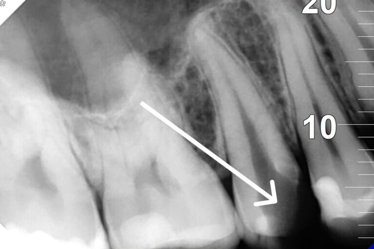
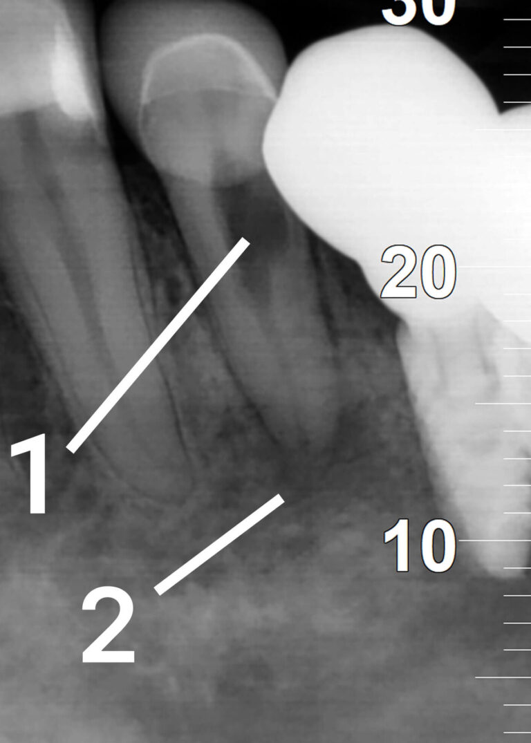
Before
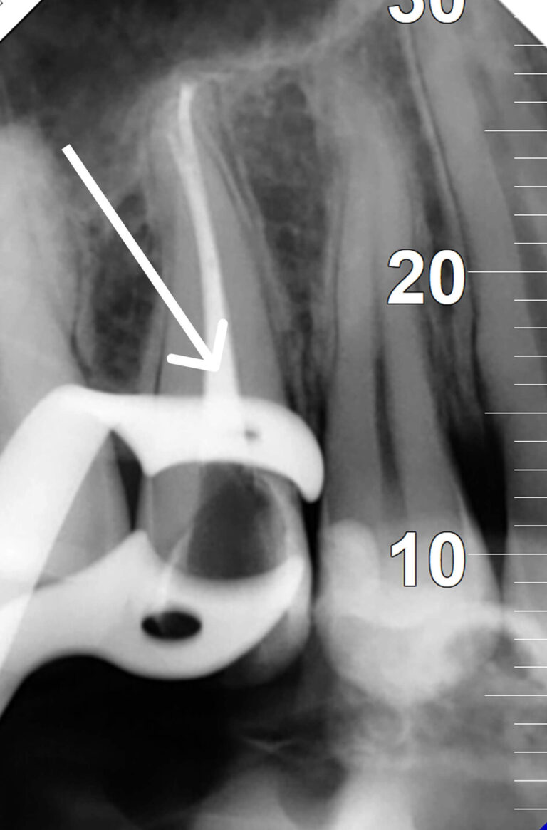
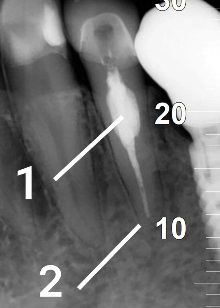
After

In brief: at KES, an exacerbation of chronic apical periodontitis of tooth 24 (upper first premolar) was treated in a single visit — chemo-mechanical cleaning and a tight obturation of the root canal, followed by a direct composite crown restoration. Single-visit endodontics is as effective as multi-visit treatment: randomized trials report ~88–97% healing at 2 years when the protocol is followed.
Key facts
| Tooth | 24 — upper first premolar (usually 1–2 canals) |
| Complaint | Constant pain that worsens on biting |
| Cause | Untreated deep caries communicating with the canal |
| Diagnosis | Exacerbation of chronic apical periodontitis + apical bone destruction |
| Treatment | Single-visit endodontics + direct restoration |
| Anaesthesia / time | Local anaesthesia, painless / 1 visit |
| Prognosis | Favourable; X-ray follow-up at 6–12 months |
What is apical periodontitis
Apical periodontitis is inflammation of the tissues around the root apex, usually of bacterial origin: infection reaches the root from a deep carious cavity through an infected or necrotic pulp. The chronic form is symptom-free for a long time and appears on an X-ray as an area of bone rarefaction at the apex (apical destruction). An exacerbation is the shift into an acute, painful phase — a typical sign being pain on biting.
Stage 1. Diagnosis
We took a periapical X-ray (CBCT when indicated), assessed the apical bone destruction and the link between the carious cavity and the canal, and ran percussion and thermal tests. Tooth 24 typically has one or two canals, which we accounted for when planning.
Stage 2. Isolation and endodontic access
The tooth was isolated with a rubber dam for sterility; infected carious tissue was removed and an access cavity to the canal orifices was made. Isolation prevents saliva and bacteria from entering the canal during treatment.
Stage 3. Chemo-mechanical cleaning
Canals were negotiated, working length was set with an apex locator, and mechanical and hand instrumentation were performed. Canals were irrigated copiously with sodium hypochlorite (NaOCl) to dissolve necrotic tissue and disinfect, and with EDTA to remove the smear layer. This is the key stage — success depends on how well the canal system is cleaned and disinfected.
Stage 4. Obturation and crown restoration
The canal was sealed three-dimensionally with gutta-percha and a sealer. In the same visit, the crown was rebuilt with a direct light-cured composite restoration, restoring shape, contact points and chewing function.
Single-visit vs multi-visit endodontics: which to choose
| Parameter | Single-visit | Two+ visits |
| Healing at 2 years | ~96% (RCT) | ~89% (RCT); difference not statistically significant |
| Indications | Patent canals, no active purulent process | Complex anatomy, heavy exudate, re-treatment |
| Time / visits | One appointment | Two or more, with a temporary dressing |
| Inter-visit re-infection risk | None | Possible if the dressing seal is broken |
Result and prognosis
The inflammation at the apex was eliminated, the canal was hermetically sealed, and the crown was restored — the tooth was preserved without extraction. The prognosis is favourable: systematic reviews and RCTs show that high-quality endodontic treatment heals the periapical tissues in most cases, and single-visit treatment is comparable in success to multi-visit. Bone at the apex recovers gradually, so a follow-up X-ray is scheduled at 6–12 months.
FAQ
Q: How many canals does tooth 24 have?
A: The upper first premolar (tooth 24) most often has one or two root canals, less commonly three. The exact number is determined by X-ray/CBCT and during treatment under a rubber dam.
Q: Can apical periodontitis be treated in one visit?
A: Yes, in most uncomplicated cases. If the canals are patent and there is no active purulent process, cleaning, obturation and restoration are done in a single appointment. RCTs show comparable success to a two-visit approach.
Q: Does periodontitis mean the tooth must be extracted?
A: In most cases the tooth is saved with endodontic treatment. Extraction is considered only with severe root destruction, a vertical fracture, or when the canal cannot be cleaned properly.
Q: Is root canal treatment painful?
A: Treatment is performed under local anaesthesia and is painless. Mild tenderness on biting for the first few days is a normal reaction that gradually subsides.
Q: Is a crown needed after canal treatment?
A: Posterior teeth (premolars and molars) are often reinforced with a crown or a ceramic onlay after endodontics: a non-vital tooth is more brittle, and a protective restoration reduces the fracture risk.
Q: What happens if periodontitis is left untreated?
A: The inflammation destroys the bone around the root, can develop into a cyst or a purulent abscess, and may lead to tooth loss and complications for neighbouring teeth.
Q: How do I know the treatment was successful?
A: Pain and discomfort resolve, and a follow-up X-ray at 6–12 months shows restored bone pattern at the root apex.
Clinic of Aesthetic Dentistry (KES), Kyiv — established in 2000; treatment is performed by top-category doctors. This material is informational; the exact plan is defined after an in-person consultation and diagnostics (X-ray/CBCT).

 Our team
 Reviews
 Blog
Blog News
News Partners
 FAQ
FAQ Vacancies
 Dental Consultation
 Tooth Decay Treatment
 Root canal treatment under a microscope
 Orthodontics
 Dental Implants
 Dental Prosthetics
 Professional Oral Hygiene
 Tooth Filling
 Periodontal disease treatment
 Aesthetic Dental Restoration
 Teeth extraction
 Surgical dentistry
 Sedation dentistry (sleep dentistry)