(1,592)


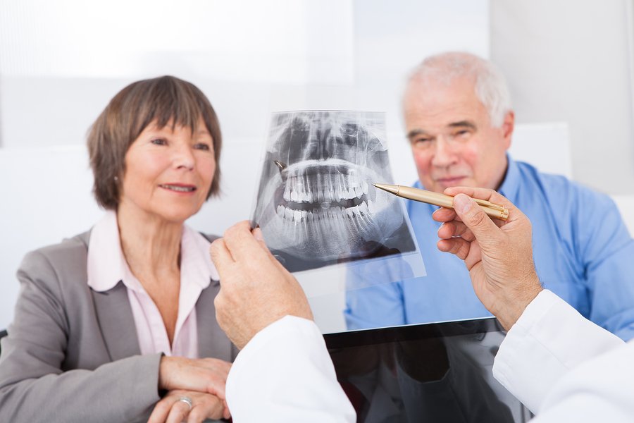
Removing the nerve of a tooth in modern dentistry is a painless and safe procedure. Its aim is to prevent the development of the inflammatory process and infectious diseases in the oral cavity.
What does a dental nerve look like in a photo? When is such treatment essential? How do they perform the nerve removal? What happens to the tooth after the pulp is removed? We answer these questions in detail below.
What Does a Dental Nerve Look Like?
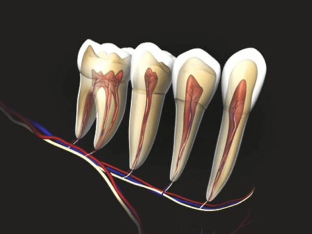
What does a tooth nerve look like? Each element of the dentition consists not only of bone tissue. A pulp containing a bundle of nerve endings and blood vessels is located at the root canal. When the nerve is irritated, sharp and unbearable ache occurs. To understand why discomfort occurs, you can study the photo of the tooth’s nerve, structure, and function.
Functions of the tooth nerve:
- Sensory perception. Pain occurs with the development of the pathological process, tissue damage.
- Nutritious. The pulp nourishes the hard and soft dentition tissues.
- Immune. Performs protective functions.
- Nerve endings are involved in dentin and enamel mineralisation.

Nerve removal in dentistry means pulp removal. In the photo, you can see the tooth nerve. This neurovascular plexus, which has a complex structure, is located inside the crown and root of the tooth. The pulp reacts to cold and hot stimuli and prevents the pathogenic microorganisms from penetrating.
Indications for Tooth Nerve Removal
Why is a nerve removed from a tooth? The main indication for removal of the tooth nerve is pulpitis. Pathology is a consequence of neglected cavities. The destroyed enamel leads to hole formation, the nerve gets exposed, it is affected by various external factors, and subsequently, pain occurs.
Complete removal of the nerve is necessary in the following cases:
- pulpitis progresses into periodontal disease;
- the presence of deep carious lesions;
- the crown gets destructed due to mechanical damage;
- the infection spreads through the apex;
- if a crown needs to be placed on a severely damaged tooth;
- severe, prolonged toothache.
How Do Dentists Remove Tooth Nerves?
How is a nerve removed from a tooth? Previously, arsenic was used for nerve extraction; the procedure took long and was dangerous and painful. However, modern endodontic treatment methods carry out depulpation at a time absolutely painlessly. Before the procedure, the doctor will tell you how the nerve is removed so that the patient understands that severe discomfort is not the case.
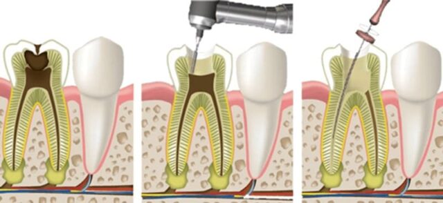
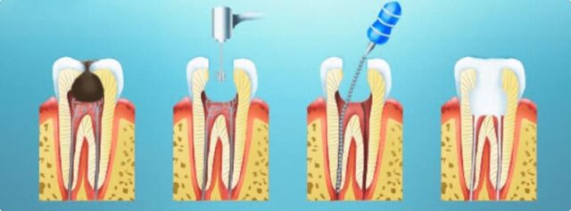
What is the procedure for dental nerve removal:
- A check-up and an x-ray. In the picture, you can see what the dental nerve looks like, its length, the presence of root canal branches, and find out to what depth the filling should be.
- The administration of an anaesthetic.
- Removal of affected tissues for pulp access.
- Pulp extraction.
- After the removal of the nerve, the dental canals and the tooth are filled. Sometimes the doctor puts a temporary filling for safety to ensure the procedure goes without complications. The dentist will put a permanent filling in a few days if there are no problems.
- Subsequent x-ray to assess the result of treatment.
If there are no complications, the removal of the tooth nerve lasts for about an hour. Many patients believe removing the nerve is painful, but this is not. Modern dentistry has everything you need to make the procedure as comfortable as possible.
What Happens to Teeth after Nerve Removal?
Understanding how the nerve is removed from the tooth and what such an intervention can cause is necessary.
After the nerve removal, the blood supply and mineralisation of the tooth stop; this dentition element is often called “dead”. Tooth enamel gets dull and appears darker; the lack of the pulp chamber cap leads to chips or tooth wall damage. With partial removal of the nerve, the tooth is destroyed more slowly.

However, you need to remove the tooth nerve. This will help to eliminate severe toothache and to avoid implant placement. Even “dead” teeth can last long if you practice good oral hygiene. Modern techniques and materials enable avoiding enamel staining and other negative consequences. It is better to restore the pulpless tooth with a crown in case of significant tissue destruction.
To prolong the life of your teeth, you need to give up sugary carbonated drinks, citrus juices, and food that can be hard on your teeth. You need to eat more food that contains iron, calcium, and phosphorus. Do not use whitening toothpaste, and practice good oral hygiene.

Tooth pain after nerve removal
In some patients, pain after removing the dental nerve bothers them for a long time. However, it is essential to understand what causes discomfort – improper treatment or physiological reasons.
Temporary pain in a pulpless tooth is normal. After active intervention in the structure of the dentition element, it takes time for the damaged tissues to heal.
In such situations, discomfort occurs with strong closure of the jaws, chewing solid food, and exposure to cold or hot stimuli. To eliminate discomfort, the doctor will prescribe painkillers and rinse solutions.
Dangerous symptoms:
- pain does not decrease or increase;
- a throbbing and stabbing tooth pain appears around the damaged tooth, especially at night;
- there is swelling of the gums and cheeks and bad breath.
If at least one such sign appears, it is necessary to urgently consult a dentist and undergo an examination to identify the cause of discomfort. This may be incomplete removal of the tooth nerve, poorly cleaned canals, or an allergic reaction to filling materials.
Do not postpone a visit to the doctor, and don’t self-medicate. This can lead to an acute inflammatory process and tooth loss.